3.2.2 IPOM (laparoscopic)
The IPOM (Intraperitoneal Onlay Mesh) technique is a special repair procedure where a mesh is introduced into the abdominal cavity and placed from the inside over the hernia opening.
To conduct this endoscopic IPOM technique, a small incision is first made in the scar-free abdominal wall [Fig. 26]. Via this approach, gas is blown into the abdomen to assure better visibility. Then an instrument with a small camera is introduced. Using this optical facility, the surgeon has a clear view on a monitor of the surgical field and the various procedural steps. Via two further small incisions, two more working instruments are advanced into the abdominal cavity. Now the surgeon can, if necessary, detach any adhesions and then expose the contents of the hernial sac [Fig. 27]. Once the hernia opening is fully exposed, a decision must be taken as to whether a synthetic mesh can be placed directly over the defect – something that is possible for defects measuring up to 8 cm – or whether the defect must be additionally constricted with a suture so that the synthetic mesh is better supported [Fig. 28]. Then the synthetic mesh, which is fitted with several sutures, is introduced and unfolded over the defect in the abdominal wall. Using a special instrument, the exposed double threads are guided out via tiny skin punctures through the abdominal wall and knotted via the small skin punctures to the abdominal wall fascia. This provides for secure fixation of the synthetic mesh to the abdominal wall [Fig. 29]. In addition, the synthetic mesh is secured from the inside by means of special tacks with titanium spirals or absorbable screws [Fig. 30]. For very big defects measuring more than 8 cm, first closing the defect with a suture provides greater support to the mesh [Fig. 31].
The endoscopic IPOM technique can only be carried out under general anaesthesia.
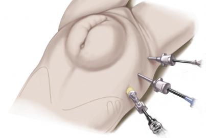
Fig. 26: Abdominal wall incision (lap. IPOM)
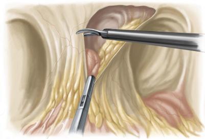
Fig. 27: Exposure of hernial sac (lap. IPOM)
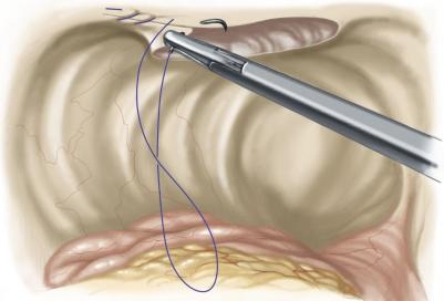
Fig. 28: Defect constriction with suture (lap. IPOM)
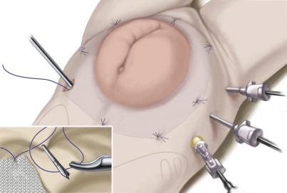
Fig. 29: Fixing synthetic mesh to abdominal wall (lap. IPOM)
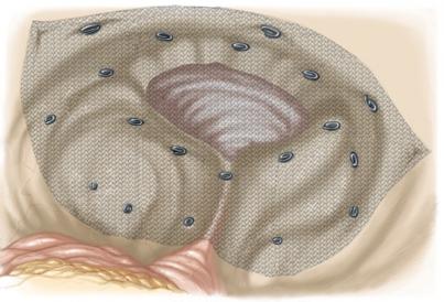
Fig. 30: Additional fixation with tacks (lap. IPOM)
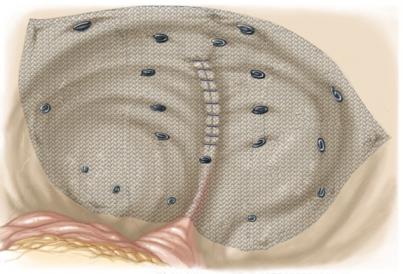
Fig. 31: Additional defect constriction with suture (lap. IPOM)